Highest reported cure rate
Recognized as the skin cancer treatment with the highest reported cure rate.
Winter Garden · Apopka · The Villages · FID Cosmetic Center
The skin cancer treatment with the highest reported cure rate.
Inside the procedure
Hearing that a biopsy shows skin cancer raises immediate questions. In this short clip our team walks through how a diagnosis is reached and how Mohs micrographic surgery is used to remove the cancer while sparing healthy tissue.
Understanding the sequence ahead of time makes the day of surgery far less daunting.
Overview
Mohs micrographic surgery is a specialized, highly effective technique for the removal of skin cancer.
What it is
Mohs surgery is typically reserved for treatment of cancers that are large, recurrent, aggressive histologically and at high risk for recurrence or metastasis. It is also indicated for cancers located in areas such as the nose, ears, eyelids, lips, hairline, hands, feet and genitals, in which maximal preservation of healthy tissue is critical for cosmetic or functional purposes.
The procedure was developed in the 1930s by a general surgeon, Dr. Frederic Mohs, at the University of Wisconsin and is now practiced throughout the world.
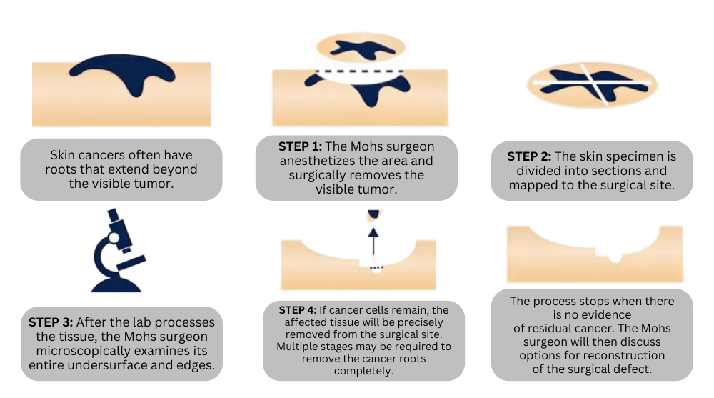
Mohs surgery differs from other skin cancer treatments in that it permits the immediate and complete microscopic examination of the removed cancerous tissue, so that all “roots” and extensions of the cancer can be eradicated. Due to the methodical manner in which tissue is removed and examined, Mohs surgery has been recognized as the skin cancer treatment with the highest reported cure rate.
How Mohs works
Skin cancers often have roots that extend beyond the visible tumor. Mohs surgery removes the cancer in stages, examining the tissue under the microscope between each stage until no cancer remains.
Conditions & indications
Large skin cancers
Recurrent skin cancers
Histologically aggressive tumors
Tumors at high risk for recurrence or metastasis
Cancers on the nose, ears, eyelids, lips or hairline
Cancers on the hands, feet or genitals
What to expect
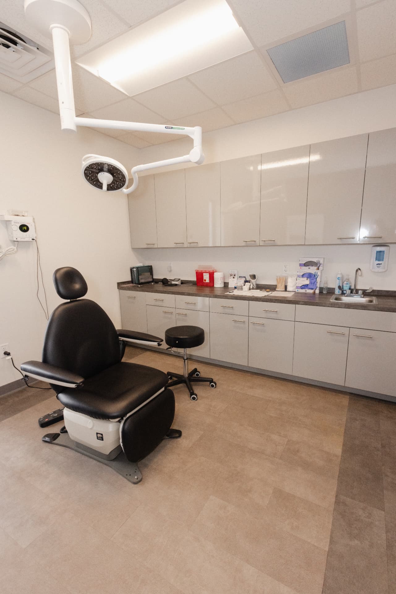
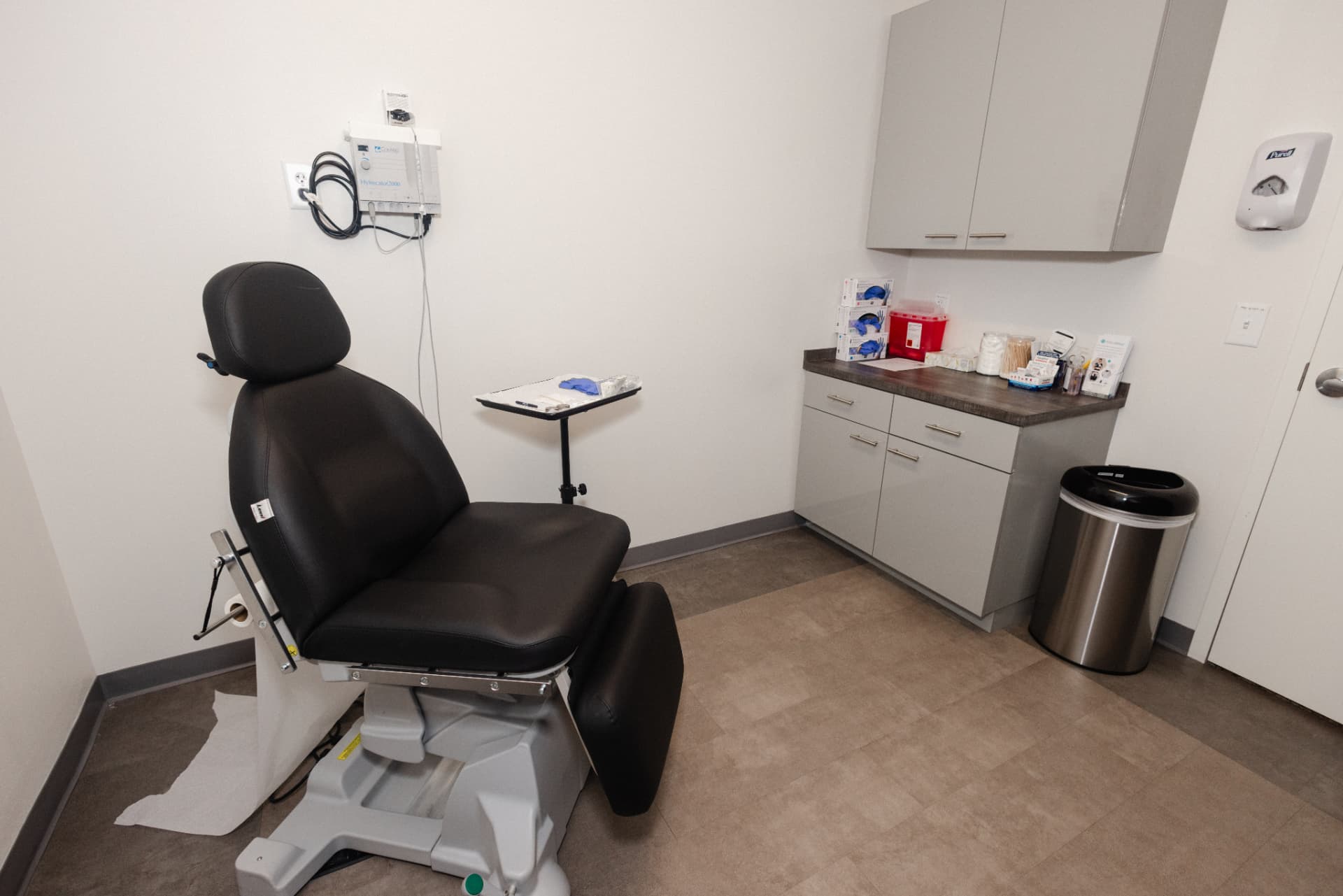
Mohs surgery is performed in our state-of-the-art office with fully equipped surgical and laboratory facilities and experienced staff.
The procedure is performed under local anesthesia.
Removed tissue is examined immediately and completely so that all roots and extensions of the cancer can be eradicated.
When clinically indicated, reconstruction of the surgical defect is part of the treatment pathway. Dr. George Papanicolaou, a double board-certified plastic surgeon, performs complex reconstruction of facial and body defects following Mohs excision of skin cancers. Detailed post-operative care is covered in our Procedural Instructions.
Before surgery
Mohs begins long before the day of surgery. Our dermatology physician assistant explains how a lesion is examined, documented and biopsied.
That evaluation determines whether Mohs is the right approach for your skin cancer.
The procedure
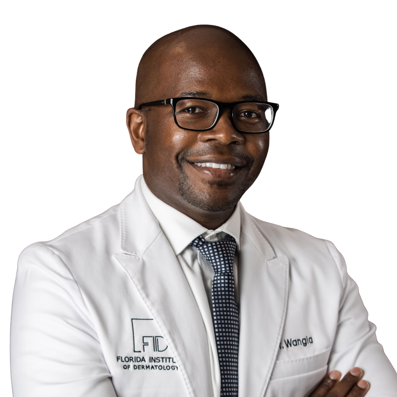
Dr. Michael Wangia is a board-certified Dermatologist, Dermatopathologist and Mohs Micrographic Surgeon. He has had an incredible level of exposure from common and rare malignant tumor pathology to challenging tumor locations and complex wound reconstruction.
When reconstruction of the surgical defect is clinically indicated, Dr. George Papanicolaou, a double board-certified plastic surgeon, performs complex reconstruction of facial and body defects following Mohs excision of skin cancers.
Benefits
Recognized as the skin cancer treatment with the highest reported cure rate.
Immediate and complete microscopic examination of the removed tissue.
Maximal preservation of healthy tissue in cosmetically and functionally critical areas.
Performed in our office under local anesthesia with our own laboratory on site.
Questions
No. Mohs surgery is performed under local anesthesia.
Recovery depends on the size and location of the surgical site and on whether reconstruction is performed. Follow the Mohs Surgery Procedural Instructions provided by your care team, which cover wound care and activity after the procedure.
We participate in most major health insurance plans, including Medicare, Blue Cross Blue Shield, Cigna, Aetna, UnitedHealthcare, Medicaid, Freedom, Optimum, Orlando Health, Multiplan and DNS (including Magellan, Devoted and ABH). Coverage depends on your specific plan, so please verify your benefits with your insurer when you schedule.
Your care team
Appointments
Book a visit at any of our four Central Florida facilities.